Content:
1. Introduction to limbic system
2. Functions of limbic system
_
Introduction to limbic system
Originally was term the limbic system used for overall nomenclature of structures separating basaly laying structures of the brain from the neocortex (word limbus means a border). Meaning of the term is widen now and in general we understand by it these structures participating on our emotional life, motivation and vegetative functions of the brain..
Functional anatomy
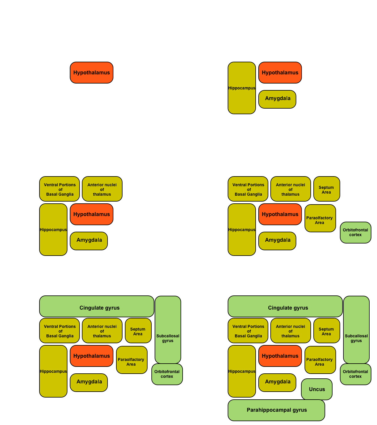
The limbic system is basicly complex mutual connected net of basal anatomic structures of the brain in which central post has the hypothalamus (anatomically and functionally). This structure is surrounded by these: the septum, parolfactory area (small region of cortex caudally from lamina terminalis), anterior nuclei of thalamus, ventral parts of basal ganglia, hippocampus and amygdala.
On the surface of the brain above those structures is located the limbic cortex consisting of orbitofrontal area (that implies position on the ventral part of the gyrus frontalis), paraterminal gyrus, cingulate gyrus and parahippocampal gyrus.
This means that at ventral and medial surface of both hemispheres is the the ring of paleocortex, which surrounds subcortical structures responsible for behaviour and emotions.
This system has important connection with the brain stem by the medial forebrain bundle, which is comprised of both ascendant and descendant fibres and interconnects areas of the septum, orbitofrontal cortex with reticular formation of the brain stem while goes through the lateral hypothalamus. Electric stimulation of this fascicle elicits very strong feeling of pleasure.
Hypothalamus
The hypothalamus has two way link with practically all of structures of the limbic system. In addition has extensive projections to:
1) Reticular formation of the brain stem
2) Higher areas of diencephalon and telencephalon
3) Across infundibulum hypothalami affects endocrine functions of hypophysis (see hypothalamic-pituitary system for more)
Despite the fact, that the hypothalamus comprises less than 1 % of the brain weight, it is crucial component, which controls the most of the vegetative and endocrine functions of the body and various aspects of our emotional behaviour.
The anatomy of hypothalamus is very complicated and various groups of nuclei and areas with specific functions were described. But our cognitive abilities are limited, because their functions we can find just through experiments which either some region stimulate or eliminate on the contrary by damaging of an appropriate anatomical defined location. On the basis of these experiments we try to assemble theories about functions of individual nuclei/areas and their mutual relations.
_
Function of limbic system
Hypothalamus
Cardiovascular regulation
Areas of the hypothalamus are able to regulate blood pressure and heartbeat frequency by neural influence very effectively.
We can generally say that stimulation of the lateral and posterior hypothalamus raises both heartbeat frequency and blood pressure. On the other hand high activity of the preoptic area decreases either of them.
This effect is arranged by the linkage of the hypothalamus with cardiovascular control centres in the reticular formation of the pons and medulla.
Regulation of body temperature
In the preoptic area are located two types of temperature sensitive neurons, perceiving the cold and perceiving the warm. Both types monitors the temperature of the blood flowing through adjacent capillaries and beyond this own receptor activity obtain informations from peripheral thermoreceptors.
If the temperature of the blood descends, then cold sensitive neurons in preoptic area rise the frequency of produced action potentials and via their projections triggers vasoconstriction of the skin and appropriate patterns of behaviour (looking for sources of the heat and the like).
On the contrary rise of the temperature stimulates warm sensitive neurons whichs projections indirectly mediate vasodilatation of the skin and respective behaviour.
Regulation of water metabolism
In the lateral hypothalamus is the area that we call the thirst center. Neurons of this structure are able to monitor the osmolarity of surrounding fluid. If there is the hyperosmolar surrounding then they shrink (by diffusion of their intracellular water to the hyperosmolar extracellular space), and this leads to the change in their activity and finally in causing the sensation of thirst.
In the supraoptic area we can find similar neurons able to monitor osmotic pressure. They raise their activity in hyperosmolar fluid and by their projections across the infundibulum to the neurohypophysis release from these axons antidiuretic hormone (ADH alias vasopressin). This hormone causes water resorption in collecting ducts in kidneys. This mechanism allows the holding of water together with preserved secretion of electrolytes and so gradual normalisation of the osmolarity. For more informations see Subchapter 7/6 and Subchapter 11/3.
Regulation of feeding
Some of hypothalamic area are closely connected with the regulation of appetite. The lateral hypothalamus is the area which causes very strong feeling of the hunger and compulsive craving to find some food (orexigenic effect). A lesion of the lateral hypothalamus leads to absolute loss of the appetite what leads to death of experimentally injured animals. According to this fact is lateral hypothalamus considered to be the hunger center.
The center of satiety is located in the ventromedial nucleus. The stimulation of this center leads to the complete loss of interest about the nourishment in experimental animals (anorexigenic effect). Lesion of this center leads to loss of the fullness feeling and in experimental animals causes morbid obesity.
It is important to remark, that there are studies which does not proved hyperphagia caused even by very precious lesions of the ventrolateral hypothalami. Because of this is its role discussed.
Corpora mammillaria are involved in the regulation of the feeding too, they probably intervene in the activity of reflexes involved in the feeding (swallowing, licking and so on).
Regulation of endocrine functions
The hypothalamus is also implicated in the regulation of the most of endocrine functions, see Subchapter 11/3 for more details.
Behavioral functions of hypothalamus and associated structures
Lateral hypothalamus
The lateral hypothalamus participates in increasing of the whole activity of beeing except its function as the center of hunger and thirst. We consider these activities are connected, because the higher activity of lateral hypothalamus leads to both feeling of the hunger and thirst and to triggering of behavioral patterns of active looking for some nourishment and water.
Strong activation manifests as an anger and high aggression.
Bilateral lesion leads to increased passivity which presents as the loss of the most of instincts.
Ventromedial nucleus
The stimulation of the ventromedial nucleus and surrounding areas leads to inverse effects than in stimulating of the lateral hypothalamus. Experimental animal has reduced appetite and behaves abnormally tame.
If ventromedial nucleus is bilaterally destroyed, then this animal starts to be hyperactive, aggressive and we can see repeated torrents of anger even after a merest provocation.
Periventricular nuclei
The stimulation of this thin group of nuclei in the near of the third ventricle leads to feeling of fear and they work as stress centres (potent motivation apparatus, see below).
Anterior and posterior hypothalamus
Anterior and posterior parts of the hypothalamus are responsible for the stimulation of the sexual behaviour, which can attain of obscure forms after the strong stimulation. Experimental animals are forced even to sexual stimulation by inanimate objects by this stimulation.
It is important to emphasize that sexual behaviour is regulated by the most of parts of the limbic system.
Amygdala
The amygdala is complex of several small nuclei located under the surface of temporal lobe cortex. It has various bidirectional connections to the hypothalamus and to many of limbic system parts. It offers various afferents from sensoric and associational areas of the cortex, in addition. Thanks to these attributes we can poetically say, that it is window by which is limbic system watching the outer world.
It processes these sensory stimuli and sends:
1) back to appropriate cortex areas
2) to the hippocampus
3) to the thalamus
4) to the hypothalamus
So stimulation of the amygdala leads to many consequences overlapping with the hypothalamus stimulation. Depending on the location of this stimulation and its intensity then raises the heartbeat frequency, pressure, starts defecation or urination and stimulates or inhibits secretion of hypothalamo-hypophyseal system hormones. Beside these effects then provokes involuntary movements (tonic moves of head, cyclic moves of limbs, sometimes even clonic tugs or chewing).
Stimulation is also able to elicit bliss or wrath and fear in experimental animal. Other parts of amygdala causes erection, ejaculation, copulatory movements, contractions of the uterus or ovulation.
Bilateral lesion of the amygdala causes Kluver – Bucy syndrome manifesting as:
1) absolute loss of the vision
2) raised curiosity and tendency to explore neighbourhood
3) quick forgetting
4) tendendency to put anything to the mouth and to swallow even solid objects
5) extreme sexual desire characterized by attempt to copulate with youngs, other species or even inanimate objects
In human manifests as in case of mentioned experimental animal, but it is very rare situation.
We can say, that function of the amygdala is to assure for the limbic system informations about environmental conditions in which is this organism situated and to arrange appropriate behavioral patterns in those situation.
Limbic cortex
The limbic cortex is the fewest discovered part of the limbic system. We think that its most important function is to convey informations from the neocortex to subcortical limbic structures and the other way around. So it is associational area of the behaviour.
Tries to stimulate the limbic cortex does not prove any convincing results to able to evaluate its function.
Experiments based on ablation of various areas of this cortex produced much more better outcomes. Removing of the orbitofrontal cortex caused development of the insomnia and motor restlessness. Removing of the gyrus cinguli fully develops the impact of stress centers and so experimental animals were much more aggressive.
Motivation
One of the most important roles of the limbic system is the function to motivate, which maintains demanded behavioral patterns and suppresses patterns which are disadvantageous or detrimental. This system forms the personality, habits and instincts of each person. Basically anything we do is in some form influenced by this motivation system.
It was discovered, that various structures of the limbic system assigns the affective component to sensory impulses (so called affective quality of the stimulus). This system determines if some stimulus is for us pleasant or not.
We divide affective qualities to two categories: satisfaction or aversion alias reward or punishment. In each of these two categories we can differentiate this affect quantitatively, this means how much is this stimulus pleasant or unpleasant.
If we stimulate some parts of the limbic system of some animal and we will cause feelings from satisfaction up to euphory we call these structures reward centers. On the other hand stimulation of other locations provoking fear, pain, escape tries we consider these centers as punishment centers.
Reward centers
They are located mostly in the neighbourhood of the medial forebrain bundle and there chiefly in lateral and ventromedial hypothalamus.
Many scientists believed that these structures are the most important punishment centers because their strong stimulation elicits anger. Today we are sure that the lateral hypothalamus is one of the most potent reward centers. In discovering these structures were find important phenomenon which we can verify in the most of these centers: after weak stimulation are induced pleasure feelings of intensity rising with the strength of the electrical stimulus, but just to some “threshold”. After the reaching of this threshold mentioned pleasure changes to this aforesaid intensive punishment, often manifesting as an anger. Every center of reward has potential ability to work as the anger center, how important is this fact is a topic of many discussions.
Other less important reward centers we can find in the septum, amygdala, part of ventral portion of basal ganglia and in the tegmentum and mesencephalon too.
Punishment centers
One of the most potent centers we can find in periaqueductal gray matter in the near of the aquaeductus Sylvii in the mesencephalon, and it continues upward as the periventricular portion of the hypothalamus and thalamus.
Other probably secondary punishment centers lies in the amygdala and hippocampus.
Simultaneous stimulation of the reward center and punishment center is often the function of reward center completely suppressed, what is substantial fact.
Role of motivation system in learning
From experiments follows that if any activity does not activate neither punishment nor reward centers, it will not be probably saved in the memory. Processing of new stimulus locates on many places of the cortex simultaneously, but when it repeats and has no affective component it losses response to this stimulus and cortex areas will be almost no activated by it. This phenomenon is called habituation. Simply, habituation is vanishing of response to repeated stimulus without affective component.
If some sensory stimulus has assigned affective component – whether is reward or punishment – and this stimulus is repeated, we will see increasing intensity of response in the contrary. Response was reinforced, we say.
Informations from our environs are selected by this mechanism and so just small part of them is saved in our memory. More than 99 % of impulses are ignored thanks to this mechanism. There is induced response to less than 1 % and only this part is determined to retention.
Subchapter Author: Patrik Maďa

