Content:
1. Functional morphology of the endocrine system
_
Functional morphology of the endocrine system
The endocrine system produces various substances called hormones that participate in coordination and control of many organs in the body. Hormones are specific biological substances secreted by endocrine cells into the intercellular connective tissue spaces and then pass into the bloodstream. Hormones affect distant organs in the body.
The endocrine system includes pituitary gland (hypophysis), pineal gland (epiphysis), thyroid gland, parathyroid glands, adrenal glands, endocrine pancreas, kidneys and gonads.
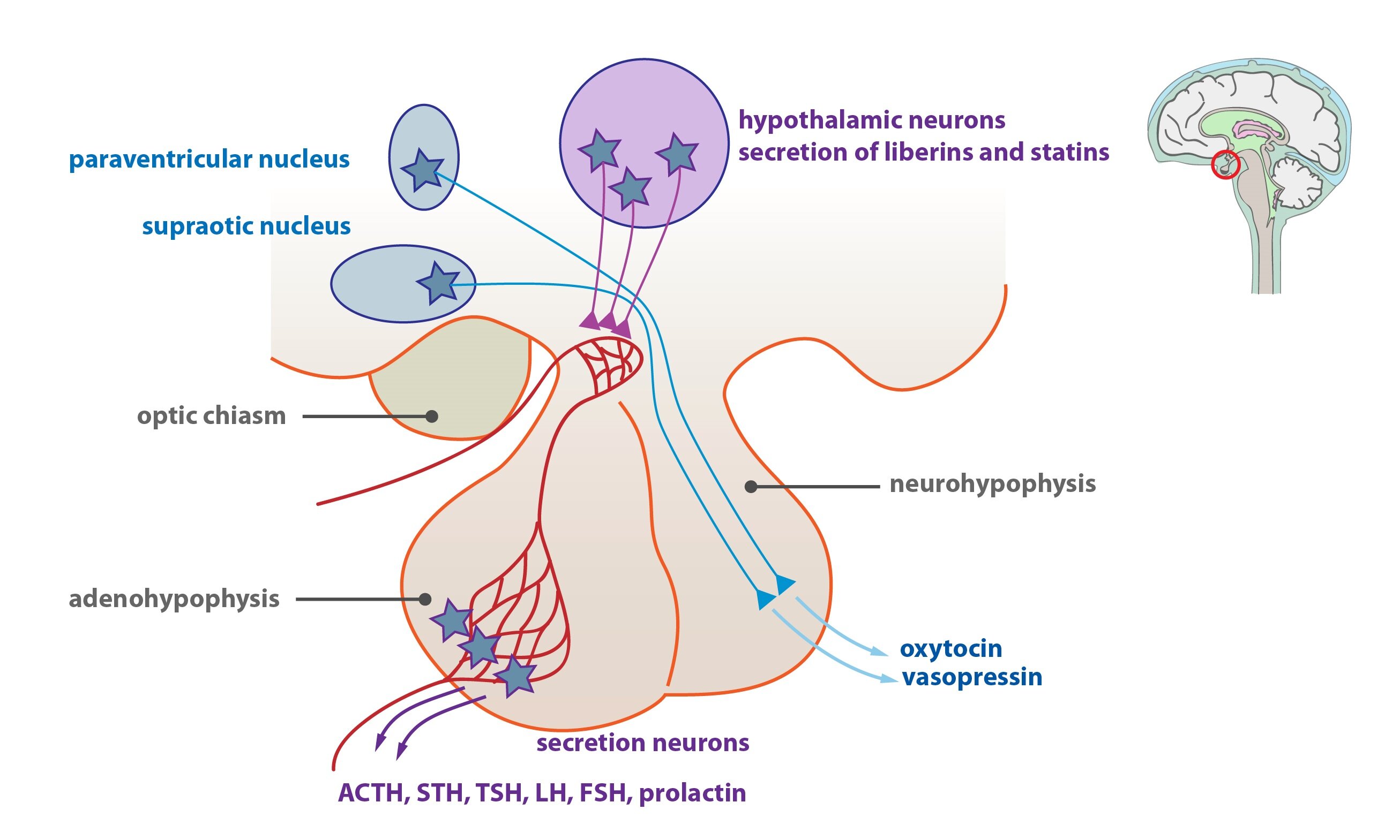
Pituitary gland (Hypophysis)
The hypophysis (pituitary gland) is embedded in sella turcica of the sphenoid bone. The hypophysis composes of adenohypophysis (anterior lobe) and neurohypophysis (posterior lobe) joined together by the infundibulum (stalk).
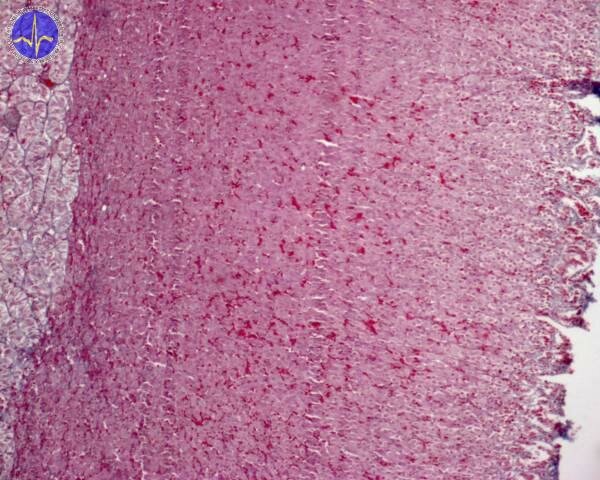
Adenohypophysis (anterior lobe)
The anterior lobe is divided into pars dorsalis, pars intermedia and pars tuberalis. The pars distalis contains a blood capillary network, chromaffin cells and chromophobe cells. Two types of chromaffin cells can be distinguished – the acidophil cells are stained with eosin and they secrete low molecular weight proteins, and the basophil cells which are stained with hematoxylin and produce glycoproteins.
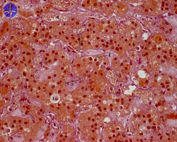
1 – Chromophobe cell, 2 – Acidophile cells, 3 – Basophils, 4 – Capillary
The acidophil cells are arranged on the periphery of the pars distalis and include somatotropic cells producing somatotropin (growth hormone) and lactotropic cells (PRL cells) producing luteotrophic hormone (also called prolactin).
The basophil cells are usually larger than acidophil cells and they include thyrotropic cells, gonadotropic cells producing follicle-stimulating hormone (FSH) and luteinizing hormone (LH) and corticotropic cells producing adrenocorticotropic hormone (ACTH). The luteinizing hormone affects the development of corpus luteum and testosterone production by Leydig cells.
Neurohypophysis (posterior lobe)
The posterior lobe contains unmyelinated axons of neurosecretory neurons, pituicytes and fenestrated capillaries. Bodies of the neurosecretory neurons are located in the hypothalamus – specifically in supraoptic nuclei (production of vasopressin which affects collecting ducts in kidneys) and paraventricular nuclei (production of oxytocin stimulating uterine contractions) of the hypothalamus.
Pineal gland (Epiphysis)
The pineal gland is located on the roof of diencephalon. Its surface is covered by capsule which continues to the pia mater. The pineal gland contains pinealocytes producing melatonin and astrocytes. The pineal secretory activity fluctuate during day depending on circadian changes of darkness and light.
Thyroid gland
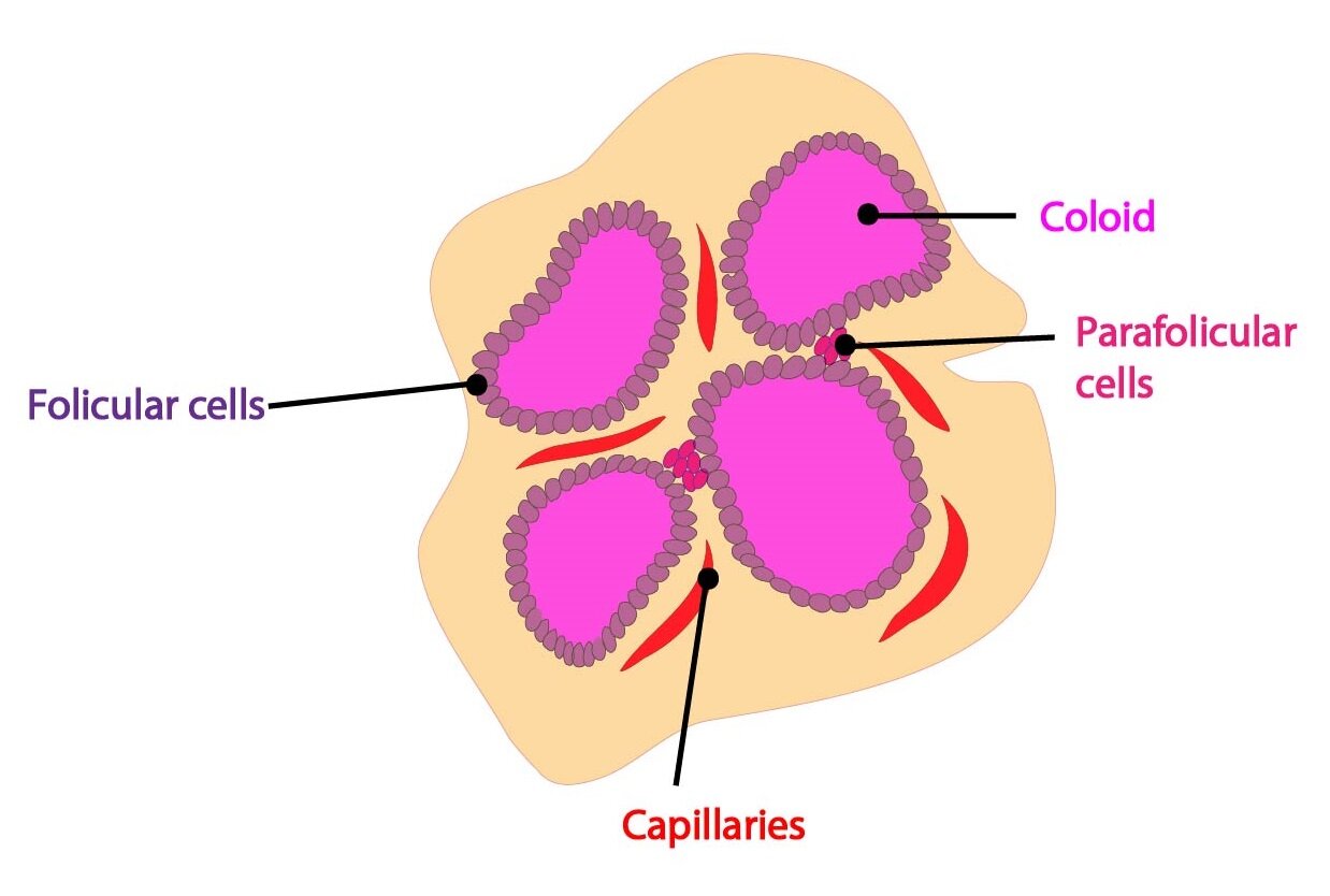
The thyroid gland is composed of two lobes connected by an isthmus. A thin connective tissue capsule surrounds the gland and sends septa extending into the glandular parenchyma. The gland contains follicular cells arranged into the follicles filled with colloid and parafollicular cells containing hormone calcitonin which decreases the blood level of calcium ions.
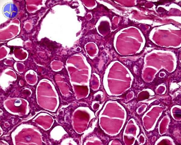
Parathyroid glands
The parathyroid glands contain primarily principal (chief) cells producing the parathyroid hormone (PTH) and also oxyphil cells. The parathyroid hormone controls the blood level of calcium and phosphate ions, stimulates the activity of osteoclasts in bones and affects the process of resorption in kidneys.
Adrenal glands
The adrenal glands are organized into two distinct regions – the cortex and the medulla. The cortex contains cells with characteristics typical of steroid-secreting cells which store their secretory products in secretory granules. The cells are arranged into the cellular cords surrounded by sinusoids. They have centrally located spherical nuclei, highly developed sER, mitochondria with tubular cristae and lipid droplets within the cytoplasm. The adrenal cortex is divided into three zones: zona glomerulosa located under the connective tissue capsule – produces aldosterone which affects the absorption of natrium ions in the distal tubules of kidneys. The second zone is zona fasciculata producing glucocorticoids (cortisol) and small amount of androgens. The third zone is called zona reticularis composed of cells with lipofuscin in the cytoplasm. These cells secrete glucocorticoids and androgens.
The medulla is formed by cellular cords and groups of polyhedral cells surrounded by reticular connective tissue. The cells produce biogenic amines – cells contain the secretory granules with catecholamines (epinephrine, norepinephrine).
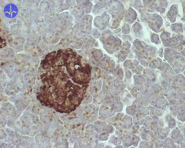
Endocrine pancreas
The pancreas is composed of exocrine and endocrine part. Exocrine pancreas is described in Chapter 9. Endocrine part is described below.
The endocrine pancreas consists of islets of Langerhans. These islets are formed by spherical cells arranged into cellular cords surrounded by network of fenestrated capillaries. Islets contain a few types of cells:
1) Beta cells –most numerous, generally located in the central portion of the islet, they secrete insulin
2) Alfa cells – located peripherally produce glucagon
3) Delta cells – produce somatostatin (inhibits insulin and glucagon secretion)
4) F-cells – produce pancreatic polypeptide
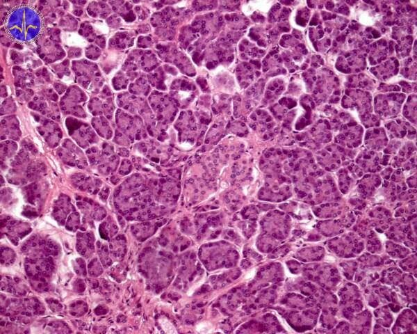
The islet is in the middle of the picture. The rest of foto is covered by exocrine acini.
Subchapter Author: Martina Šajdíková
![]()