Content:
1. Morphology of spleen
2. Function of spleen
_
Morphology of spleen
Spleen is the largest lymphatic organ. It is enclosed by a solid connective tissue capsule composed of dense collagenous connective tissue with small amount of reticular fibers and dispersed smooth muscle cells. The surface of capsule is covered by serous membrane with mesothelium. From the capsule extend connective tissue trabeculae into the parenchyma of the organ. The splenic parenchyma (splenic pulp) is divided into two functionally and morphologically different regions: white pulp and red pulp. These titles are based on the color of fresh sections of splenic parenchyme. It also contains venous sinuses. At the histological slide, a white pulp resembles spherical or elongated whitish structures surrounded by red pulp. A reticular connective tissue serves as mechanical support in spleen.
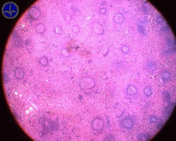
White pulp
White pulp consists of lymphatic tissue cords (mostly accumulation of lymphocytes) surrounding course of an artery in some places interrupted by lymphatic nodules (noduli lymphatici lienales). In hematoxylin-eosin stained slides, white pulp appears basophilic (because of the dense heterochromatin the nuclei of the numerous lymphocytes). Branches of the splenic artery enter the white pulp from trabeculae. Within the white pulp, these arteries are called the central arteries. Branches of the central arteries constitute a capillary network supplying the white pulp and marginal zone. The central arteries continues into the red pulp and branch into the thin straight penicillar arterioles.
Lymphocytes that aggregate around the central artery (mostly T-lymphocytes) create the periarterial lymphatic sheath (PALS). Contrarily, the lymphatic nodules are formed chiefly by B-lymphocytes. These nodule usually contains germinal center developed by proliferation of B-lymphocytes after their activation. In humans, the germinal centre develops within 24 hours after antigen exposure. These nodules are called Malpighian corpuscles (not to be confused with renal corpuscles that have the same name).

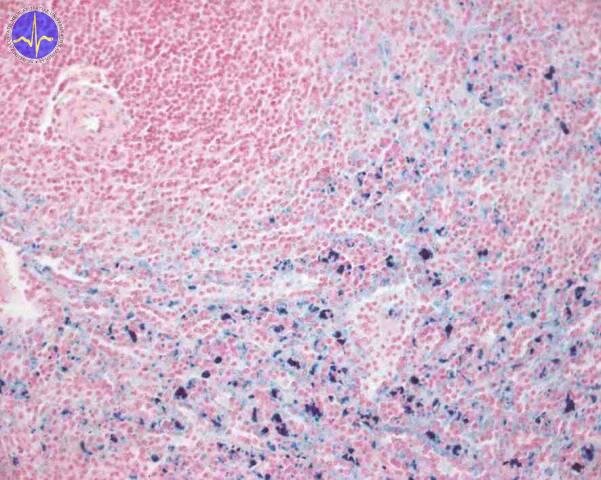
Red pulp
Red pulp consists of splenic sinuses and splenic cords (cords of Billroth) formed by reticular cells and fibers. Red pulp contains a lot of blood. In the meshwork of reticular cells and reticular fibers are lymphocytes, macrophages, large numbers of erythrocytes (normal as well as senescent), plasma cells, dendritic cells and granulocytes. The endothelium lining the splenic sinuses is fenestrated, formed by extremely long cells.
On the boundary between white pulp and red pulp, the marginal zone is created. It consists numerous actively phagocytic macrophages but relatively small amount of lymphocytes.
_
Function of spleen
Spleen is the largest lymphatic organ in a human body and it is important for many functions. The most important are proliferation and differentiation of lymphocytes and destruction of erythrocytes. It also serves as storage of blood which can be used in hypovolemia (decreased blood volume due to bleeding or long-lasting physical strain).
Proliferation and differentiation of lymphocytes
Proliferation and differentiation of lymphocytes occur in the white pulp. Then, these cells migrate through marginal zone into the red pulp venous sinusoids and are released into blood. It also plays important role in immune defence of body. It contains numerous T and B lymphocytes and also phagocytic immune cells. Its function is equivalent to the lymph nodes, but in contrast to that, spleen is classified as a part of blood vessel system (not lymphatic vessel system). Spleen removes antigens from blood and provides antigen presentation by APC’s (antigen presenting cells). Due to that, B lymphocytes are activated and they transform into antibodies (immunoglobulins) secreting plasma cells. There are also present the memory B cells which are able to rapidly activate the secretion of antibodies after exposure to the specific antigen.
Destruction of erythrocytes
The median lifetime of erythrocytes is about 120 days. During this time, erythrocytes are exposed to many adverse effects (e.g. oxidative stress) and that leads to their damage and exhaustion. Erythrocytes have no molecular biology technique to repair their own damaged proteins and these damages gradually accumulate. Subsequently, the function of erythrocytes is also affected. The role of the red pulp is primarily blood filtration and concurrently, it monitors the level of damage of erythrocytes. Spectrin, the cytoskeletal protein of erythrocytes, contributes to the shape and elastic property of these cells and allows erythrocytes to pass through the thinnest capillaries. The red pulp works as mechanical meshwork which removes senescent, damaged or abnormal erythrocytes unable to pass through thin (2-3 μm) spaces between cells of spleen sinusoids. Removed erythrocytes are broken down by phagocytes, particularly numerous in red pulp.
Iron in the spleen
Storage of blood
Spleen can release up to 100 ml of blood in case of emergency. This blood is stored in the venous sinuses and pulp. In contrast to other blood reservoir, the release of splenic reserve increases hematocrit by 1-2 %. This increase is caused by release of many erythrocytes stored in trabecular meshwork of red pulp. Release of erythrocytes is stimulated by increased activity of sympathetic nervous system which causes constriction of splenic vessels (including vascular structures in pulp) and release of red blood cells.
Subchapter Authors: Martina Šajdíková and Lucie Nováková
![]()