Content:
1. Functional Morphology of the Kidneys
_
Functional morphology of kidney
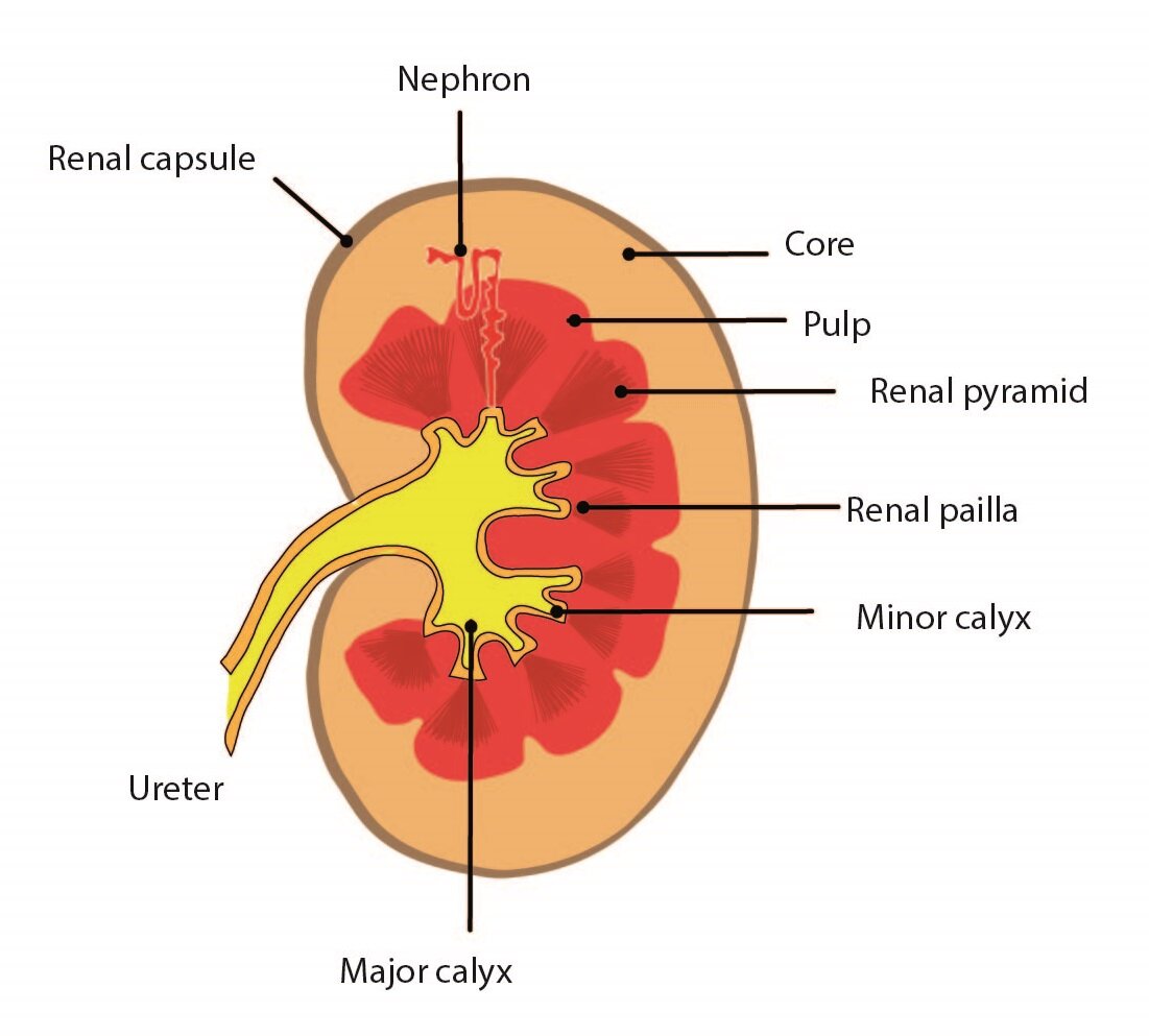
Kidneys are paired retroperitoneal bean-shaped organs weighting about 150 g. Macroscopically, kidney is distinguished into cortex with granular appearance surrounded by fibrous capsule and medulla with striated appearance. Kidneys are surrounded by adipose tissue – capsula adiposa.
Renal medulla
Medulla is composed of renal pyramids whose bases are attached to the cortical tissue and define the bordeline between cortex and medulla. The pyramidal vertices are heading to hilum and create renal papillae surrounded by smooth muscle cells allowing evacuation of urine to the renal pelvis.
Renal cortex
Cortex is about 5 to 7 cm thick. Between the renal pyramids, cortical tissue penetrates to the renal sinus and created renal columns. The cortex sends strips penetrating the medulla, called striae medullares.
Nephron
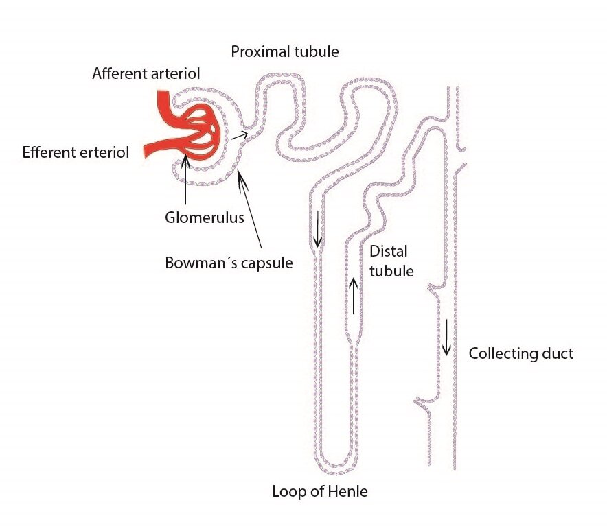
The nephron is principal structural and functional unite of kidney. Each kidney contains about 1 million nephrones. Each nephrone is composed of glomerulus, Bowman’s capsule, proximal tubule, loop of Henle, distal tubule and collecting tubules.
Renal corpuscle (Malpighi corpuscle)
The renal corpuscle is composed of glomerulus and Bowman’s capsule. Each corpuscle has two poles – vascular and urinary pole. At vascular pole, the afferent arteriole enters the glomerulus and efferent arteriole leaves it. At urinary pole, the outer parietal layer of Bowman’s capsule changes to proximal tubule.
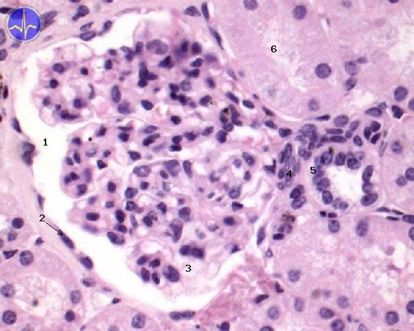
1 – Bowman’s space, 2 – Parietal epithelium of Bowman’s capsule (arrow points to the nucleus of the cell), 3 – Glomerulum, 4 – Mesangial cells in juxtaglomerular aparatus, 5 – Macula densa of the distal tubule, 6 – Proximal tubule
Glomerulus
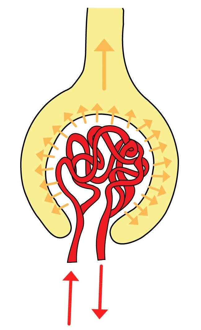 Glomerulus is composed of capillary tuft. It is surrounded by Bowman’s capsule consisting of two layers (visceral and parietal layer). The spaces between capillaries are filled with mesangium formed by intraglomerular mesangial cells and mesangial matrix. Intraglomerular mesangial cells are located near the endothelial lining of capillaries and they have angiotensine-1 receptors on their surfaces.
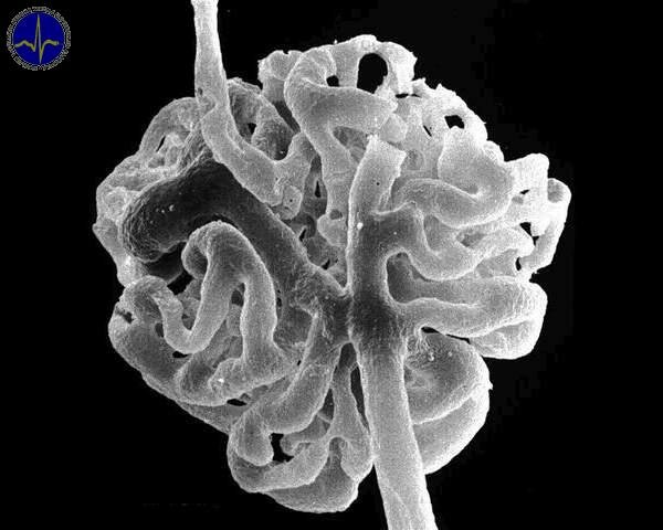
The scanning electron micrograph of glomerulus. It is formed by tuft of capillaries with afferent and efferent arterioles.
Bowman’s capsule
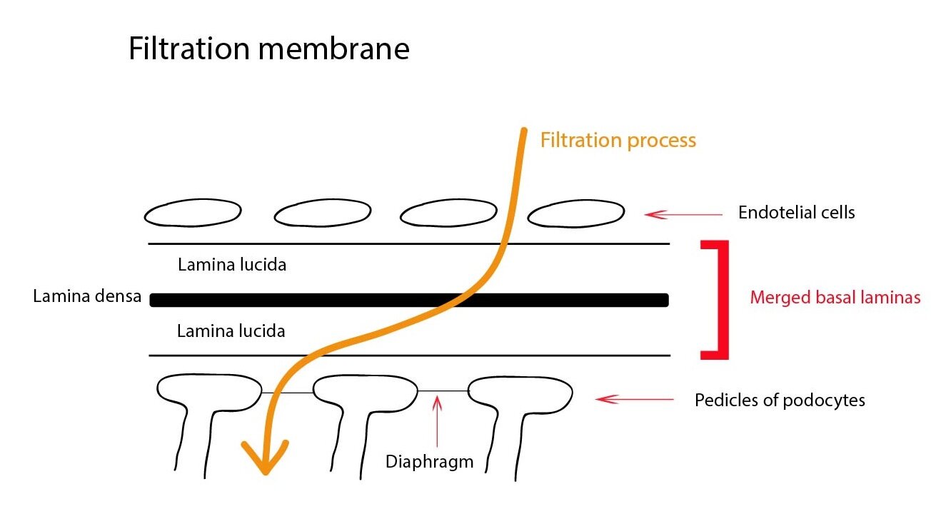
Bowman’s capsule consists of inner (visceral) layer surrounding the glomerulus and outer (parietal) layer bordering the renal corpuscle. Between these two layers is urinary space where the primary urine is produced. The parietal layer is formed by simple squamous epithelium. The visceral layer is formed by podocytes – epithelial cells with pedicles surrounding glomerular capillaries. Pedicles of adjacent podocytes interdigitate and spaces between these interdigitating processes are covered by thin slit diaphragm. These filtration slits are components of filtration barrier for for ultrafiltration of blood plasma and thus for primary urine production.
Filtration barrier
The primary urine is filtered across this barriers into the urinary space. This filtration barrier is composed of capillary endothelium, podocytes and their fused basement membranes. In an adult human, approximately 125 ml of primary urine is produced per minute. The volume of definitive urine is about 1,5 l per day.
Proximal tubule (PT)
The proximal tubule consists of two parts – tortuous (pars covoluta) and straight (pars recta). It is lined by simple cuboidal epithelium. These epithelial cells are relatively large engaged in absorption and ion transport. They exhibit the brush border, containing large amount of alkaline phosphatase, on their apical surface On the basal surface are basal striations containing numerous ion pumps. The proximal tubule is responsible for absorption of amino acids, proteins, glucose, ions, water etc.
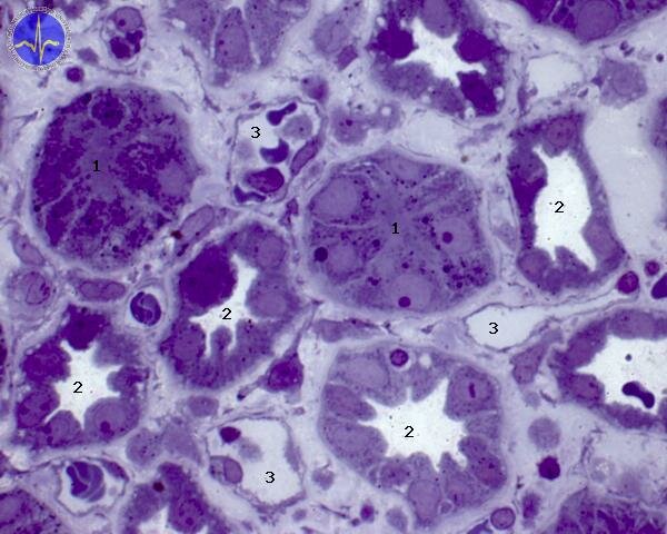
1 – Proximal tubule, 2 – Distal tubule, 3 – Peritubular kapillaries: Proximal tubule cells are larger, so on transverse section of tubule you can see less of them. Those cells are darker, because they contain more of transporting vesicles and more of mitochondrias. Lumen of proximal tubule is fullfilled with brush border. Distal tubules have free lumen (no brush border).
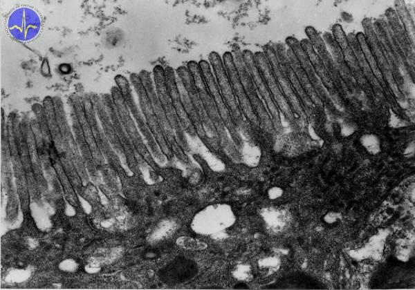
Brush border covering the apex of proximal covoluted tubule cells
Loop of Henle
This U-shaped loop consists of two limbs – the descending (thin, penetrates the medulla and is lined by simple squamous epithelium) and ascending (thick, heading back toward cortex, lined by simple cuboidal epithelium) limb. Structurally, it resembles the distal tubule. The descending limb is highly permeable to water and the ascending limb actively transports ions.
Distal tubule
The distal tubule lined by simple cuboidal cells is located in the cortex. The cells of lining epithelium are responsible for active transport of ions and they exhibit prominent basal striations. The absorption and secretion in the distal tubule is under hormonal control (e.g. aldosterone). Macula densa is an area where the wall of distal tubule closely attaches to the vascular pole of renal corpuscle.
Juxtaglomerular apparatus
The juxtaglomerular apparatus consists of macula densa and attaching part of afferent arteriole. The cells of juxtaglomerular apparatus affect blood pressure by renin production (stimulation of Renin-Angiotensin-Aldosterone axis).
Collecting tubule
The collecting tubule is lined by simple cuboidal epithelium with transition to simple columnar (as tubule increases in size). Two different types of cells are present: the darker intercalated cells with numerous mitochondria and the pale-staining principal cells which are affected by antidiuretic hormone (ADH).

1 – Principal cells, 2 – Intercalated cells (dark, secrete HCO3– and H+, reabsorb K+)
Excretory passages of urinary system
The excretory passages are composed of renal pelvis, minor calices, major calices, ureter, urinary bladder and urethra. These part have relatively similar histological structure: mucosa (tunica mucosa), smooth muscle (tunica muscularis) and adventitia (tunica adventitia):
1) Tunica mucosa – consists of epithelium (transitional epithelium) and lamina propria mucosae (loose collagenous connective tissue)
2) Tunica muscularis – smooth muscle bundles
3) Tunica adventicia – loose collagenous connective tissue
Ureter
The ureter is lined by transitional epithelium. The muscular coat is arranged into inner longitudinal and outer circular layer. The muscular coat of distal parts of ureter have the third layer – outermost longitudinal layer.
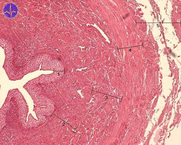
1 – Transitional epithelium, 2 – Lamina propria mucosae, 3 – Longitudinal layer of smooth muscle, 4 – Cicular layer of smooth muscle, 5 – Adventitia
Vesica urinaria (Urinary bladder)
The urinary bladder is lined by transitional epithelium (6 to 8 cellular layers). The muscular coat consists of three layers: plexiform, circular and outermost longitudinal. The middle layer forms bladder sphincter (musculus sphincter vesicae).
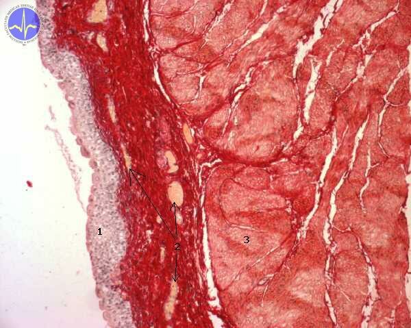
1 – Transitional epithelium, 2 – Lamina propria mucosae (arrows point to vessels), 3 – Tunica muscularis
Urethra
The proximal part of urethra is lined by transitional epithelium and the distal part is lined by stratified squamous epithelium.
Subchapter Authors: Martina Šajdíková and Lucie Nováková
![]()