Content:
1. Functional morphology of muscles
_
Functional morphology of muscles
Muscle tissue is composed of myoblasts (muscle cells) and connective tissue. The primary function of muscle tissue is contraction responsible for many types of movement in human body – e.g. movement of organs or the whole body, blood circulation etc. Muscle tissue is distinguished into the striated skeletal muscle, striated cardiac muscle and smooth muscle.
The smooth muscle develops from mesenchyme, skeletal muscle from mesodermal origins – myotomes, and cardiac muscle from splanchnopleuric mesoderm.
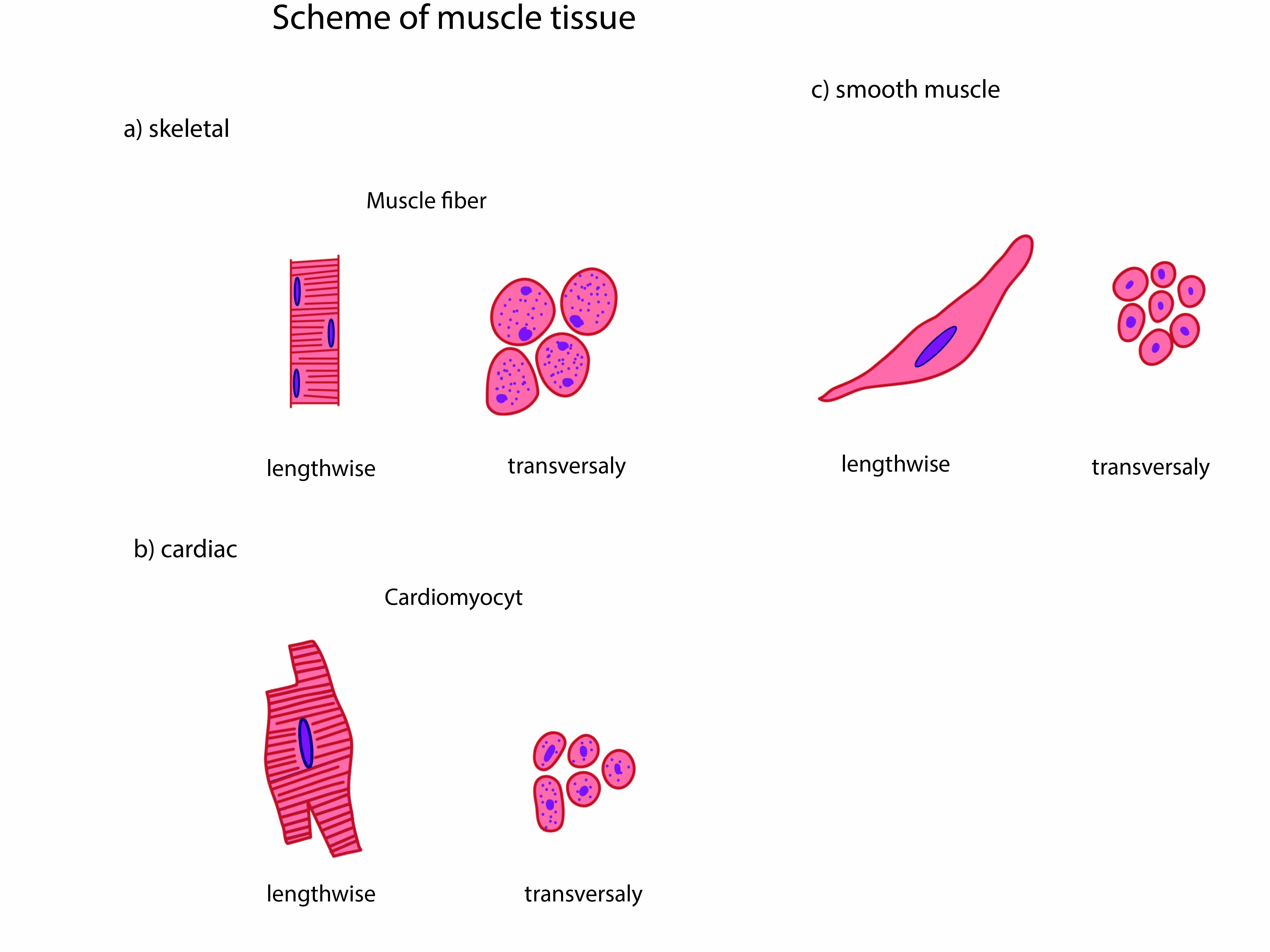
Striated skeletal muscle
The skeletal muscle consists of muscle fibers (long multinucleated syncytium) whose contraction is fast and regulated by will. Some cellular components are called differently at muscle tissue, e.g. the cytoplasm of muscular cell is called the sarcoplasm, the endoplasmic reticulum is called sarcoplasmic reticulum and cell membrane is called sarcolemma
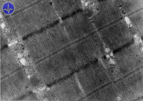
The sarcoplasm contains densely arranged longitudinally oriented myofibrils composed of two types of myofilaments – thin actin filaments and thick myosin filaments. The mutual interaction of actin and myosin enables the muscle contraction (thus these proteins are called contractile proteins). Shortening of myofibrils during the contraction is due to the sliding filament mechanism – actin filaments slide over the myosin filaments. The length of thick and thin myofilaments remains the same.
Photomicrograph of a skeletal muscle shows cross-striation – visible as alternating light and dark bands of myofilaments which differ in staining and birefringence. In polarizing microscopy, the dark bands are birefringent and better stained, they are called anisotropic A-bands (formed by thick myofilaments). The light bands are monorefringent and poorly stained, they are isotropic I-bands (created by thin myofilaments). The endings of thick and thin filaments are overlapped and due to that, on each A-band are distinguishable two peripheral segments containing both types of myofilaments and one central segment containing only myosin filaments called H-zone. I-bands are bisected by dense line called Z-line. A-bands are bisected M-line. The sarcomere is the regularly alternating portion of myofibrils between two adjacent Z-lines.
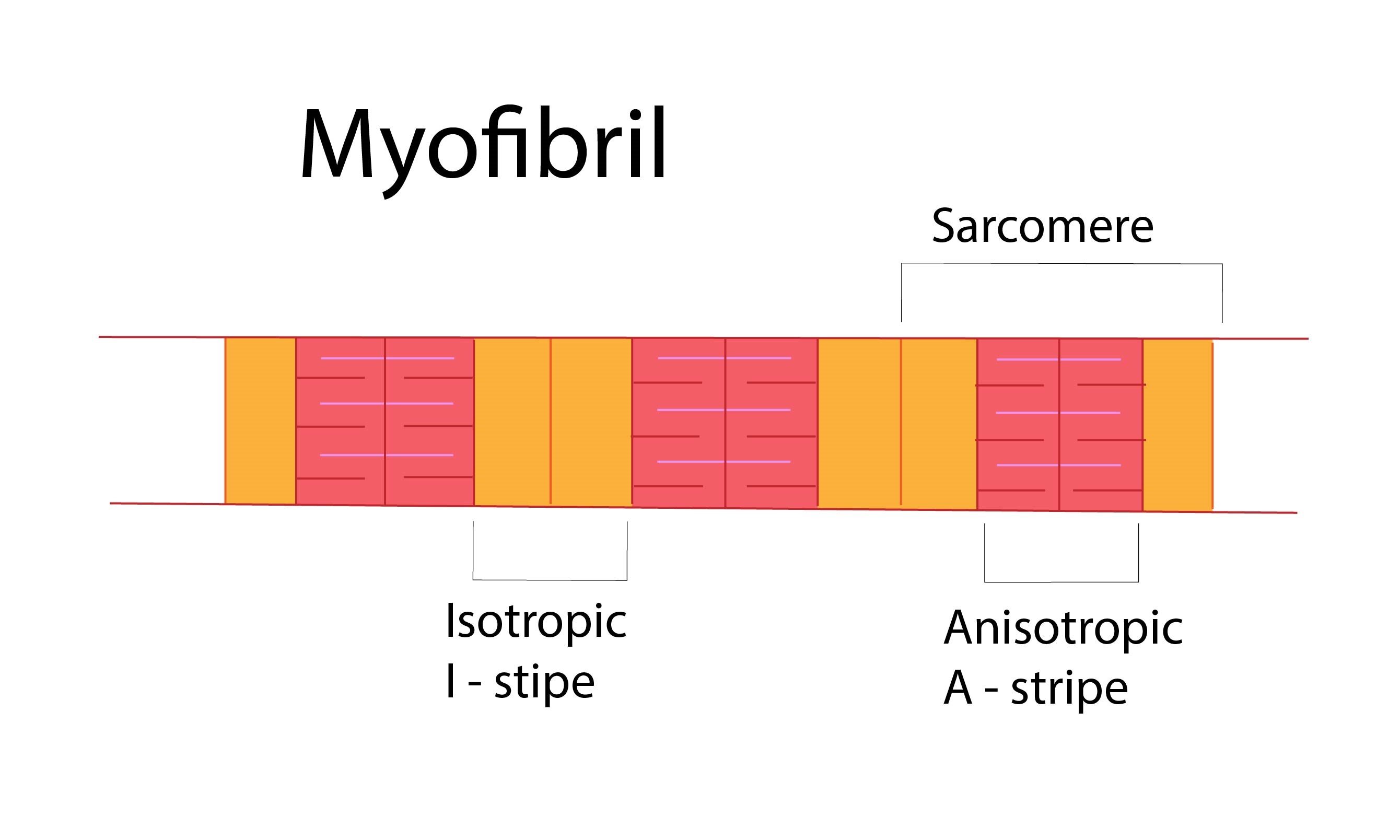
Myofibril
Striated skeletal muscle
The thick myofilaments containing myosin protein evince ATP activity and remind golf club-shape.
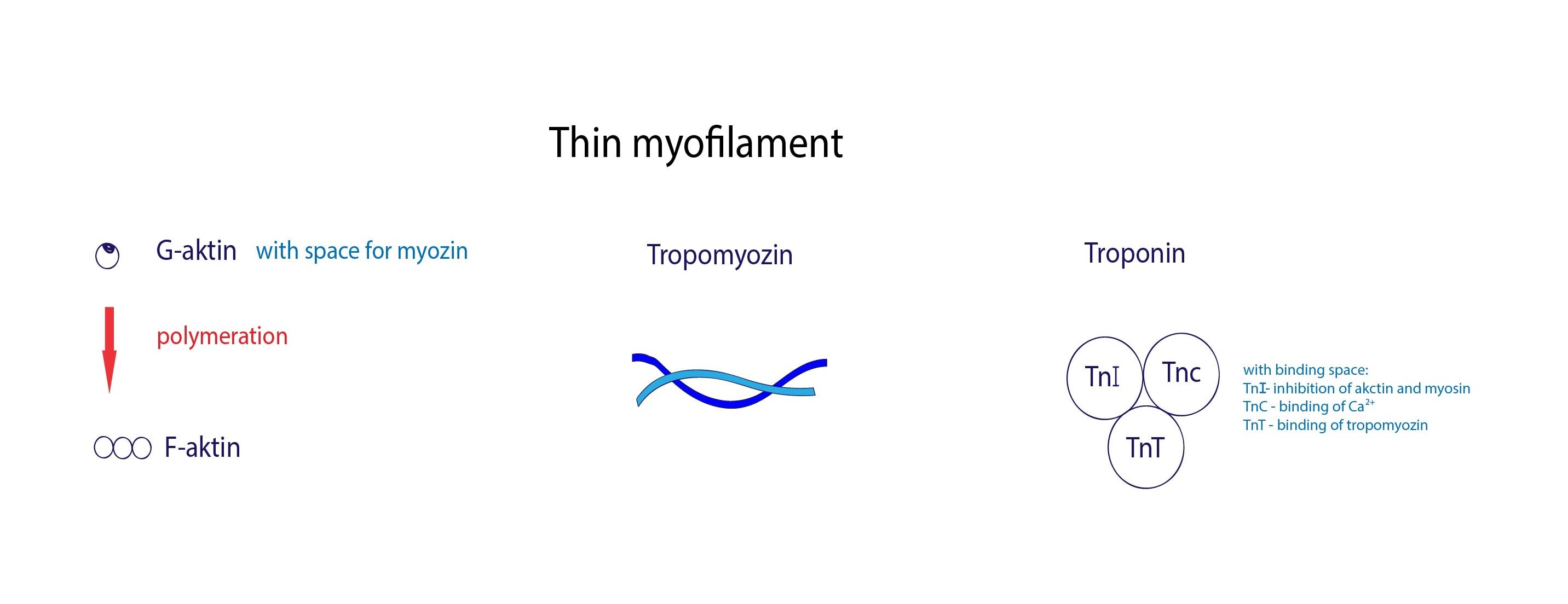
The thin filaments consist of actin and regulatory proteins tropomyosin and troponin. Actin filaments are formed by double-stranded helix of F-actin which arises from G-actin. G-actin (G means globular) polymerizes to form double-stranded helix of F-actin (F means fibrillar) and has a binding site for myosin. Tropomyosin consists of double-helix of two polypeptides and has a binding site for troponin. Troponin is a complex of three protein subunits:
1) TnT – binds to tropomyosin
2) TnC – binds Ca2+
3) TnI – inhibits actin-myosin interaction
Sarcoplasm contains also myoglobin, glycogen granules, mitochondria and oval nuclei located immediately beneath the plasma membrane.
Sarcolemma invaginates into muscle fiber and creates transverse tubular system or T-system composed of numerous T-tubules. These T-system is responsible for spreading of depolarization over the entire muscle fiber. T-tubule is localized on the boundary between A-band and I-band surrounded by adjacent sarcoplasmic reticulum cisternae – these cisternae and T-tubule form so called triad. Each sarcomere involves two triads.
Muscle fibers are classified according to the function and morphology into two groups:
The red fibers contain myoglobin and numerous mitochondria. Contraction is slower but also longer-lasting and fatigue-resistant. On the other side, white fibers contain less mitochondria and myoglobin but they are fast and fatigue-prone. Most of skeletal muscles contain both types of muscle fibers.
Individual muscle fibers are surrounded by layer of reticular connective tissue fibers called endomysium. Muscle bundles (fascicles) are surrounded by thicker connective tissue layer called perimysium (it sends septa between the muscle bundles) and a dense connective tissue sheath of muscle is called epimysium.
Cardiac striated muscle, myocardium
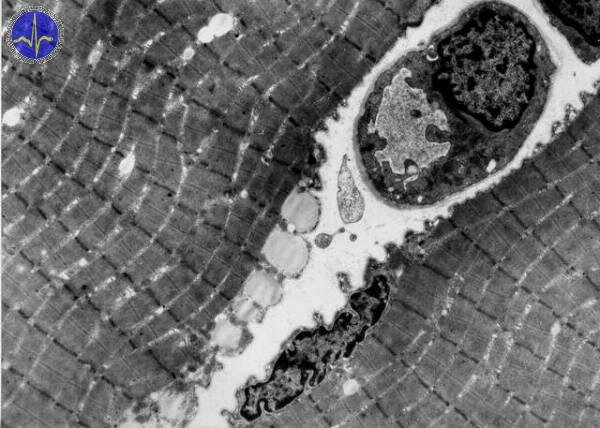
The cardiac muscle is composed of columnar cardiomyocytes containing cross-striated myofibrils in their cytoplasm. The cross-striation resembles skeletal muscle. Each cardiomyocyte contain one or two large light nuclei located centrally surrounded by numerous mitochondria. In the cytoplasm are found lipid droplets containing triacylglycerols, glycogen and lipofuscin (yellowish-brown color) granules. Photomicrograph shows prominent intercalated discs representing stepped arrangement of cardiac muscle. These intercalated discs ensure special connection between cardiomyocytes (they branch and their processes interdigitate with neighbouring cardiomyocytes). In area of intercalated disc are found fasciae adherentes (on transverse sites of discs), maculae adherentes (desmosomes) ensuring the tight connection between discs, and gap junctions allowing cellular communication.
Smooth muscle
The smooth muscle is composed of elongated spindle-shaped cells about 20 to 500 μm large (the largest are contained in uterus of pregnant woman).
The cells are present individually or in slight bundles found e.g. in walls of hollow organs (intestine, uterus etc.). The cells contain rod-shaped nuclei located centrally in the widened portion of cells. Nuclei are surrounded by numerous mitochondria, free ribosomes and Golgi complexes. Sarcoplasmic reticulum is reduced, membrane lacks T-tubules. Cytoplasm contain actin and myosin myofilaments which do not form myofibrils. Intermediate filaments are also found containing chiefly desmin and vimentin. The dense bodies are analogue of Z-lines in striated muscle and are responsible for anchoring the thin actin and intermediate filaments. These dense bodies are connected to the sarcolemma or dispersed in cytoplasm.
The smooth muscle is found in many organs of the human body – walls of hollow organs (GIT, respiratory system, urogenital system, skin, walls of vessels or iris.
Subchapter Authors: Lucie Nováková and Martina Šajdíková
![]()